
What is cutaneous polyarteritis nodosa?
Cutaneous polyarteritis nodosa (PAN) is a rare form of vasculitis(in ammation of blood vessels) that involves smalland medium-sized arteries of the dermis and subcutaneous tissue i.e. the deeper layers of the skin. It is sometimes called periarteritis nodosa.
How does it relate to systemic polyarteritis nodosa?
Although identical skin lesions are common in systemic PAN, cutaneous PAN should be considered a separate disease and distinguished from systemic PAN, as the clinical course and management of these conditions di er from each other.
PAN is a vasculitis that causes destructive in ammation of medium-sized muscular arteries of multiple systems including the liver, kidney, heart, lung, gastrointestinal tract, musculoskeletal and nervous systems. Systemic PAN is a potentially life-threatening form of vasculitis, whereas cutaneous PAN usually runs a chronic but relatively benign course.
Who gets cutaneous polyarteritis nodosa?
Cutaneous polyarteritis nodosa often starts in childhood or adolescence. It is rare.
In most cases of cutaneous PAN, the disease is triggered by certain infections, particularly Group A streptococcus, hepatitis B, hepatitis C, human immunode ciency virus,parvovirus B19 (the cause offth disease). Genetic defects lead to over-reaction to the infection.
What is the cause of cutaneous polyarteritis nodosa?
Cutaneous PAN results from a complex interaction of autoinNammatory and autoimmune factors, and immunode ciency.
Autosomal recessive mutations in the CERC1 gene have been implicated in some patients with cutaneous PAN. This genetic abnormality results in deUciency in the ADA2 protein (DADA2). DADA2 can also cause immunodePciency. ADA2 is a plasma protein essential for development of endothelial cells (these line the blood vessel wall), and leucocytes (white blood cells). DADA2 leads to uncontrolled, chronic activity of neutrophils and damages endothelial cells.
What are the clinical features of cutaneous polyarteritis nodosa?
Clinical features of cutaneous polyarteritis nodosa relate to in ammation or occlusion of small and medium-sized blood vessels in the skin and sometimes in other organs. It tends to have periods of activity and remission.
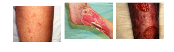
Vasculitic lesions are most often found on the legs and feet. Other areas that may be aVected include the arms, trunk, buttocks, and head and neck. They are most likely on pressure points such as the knees, back of the foot and lower leg.
- Tender lumps appear under the skin, especially on the thighs and lower legs. These usually measure between 4–15 mm in diameter and follow along the course of medium-sized arteries.
- Larger in ammatory plaques may be seen. These tend to have nodules along the edges. As the plaques heal, they leave patches of postin ammatory pigmentation.
- Infarcts in the skin present as purple or black patches or blood-Nlled blisters. They are dead areas of skin due to blocked blood vessels.
- Small vessel vasculitis maypresent as palpable purpura
- Blistering and ulceration may occur.
- Livedo reticularis mayappear (a starburst dusky discolouration).
Systemic symptoms in cutaneous polyarteritis nodosa
In addition to the skin problems, patients with cutaneous PAN may also have generalised symptoms such as malaise, fever, sore throat, and joint and muscle aches and pains. Neurological symptoms may also be present and include numbness, tingling, sensory disturbances, weakness, and absent rePexes.
How is cutaneous polyarteritis nodosa diagnosed?
Skin biopsyofa typical lesion is often performed to make an accurate diagnosis of cutaneous PAN. A specimen showing panarteritis(in ammation of all blood vessels in the skin sample) is the only deNnitive proof of PAN.
Laboratory tests of blood samples are generally unhelpful in diagnosing or monitoring cutaneous PAN, as blood counts and chemistry are often normal. They are initially required to determine the cause of vasculitisor to exclude other organ involvement as occurs in systemic PAN.
Levels of ADA2 protein and/or CERC1 gene can be measured in some centres.
What is the treatment for cutaneous polyarteritis nodosa?
As cutaneous PAN is rare, there are no randomised trials to guide treatment. Standard treatment follows guidelines for other forms of vasculitis. The mainstay of treatment is often with oral corticosteroids(prednisone).
Other active treatments for cutaneous PAN may include:
- Colchicine
- Cyclophosphamide
- Intravenous immunoglobulin
- Penicillin toprevent ares after streptococcaltonsillitis or cellulitis
Warfarin (with an international normalised ratio [INR] of 3) has been reported to be e ective in 3 cases of cutaneous polyarteritis nodosa with improvement in livedo reticularis and healing of ulcers.
New therapies are being developed for patients with cutaneous PAN that have DADA2. These unproven treatments include:
- Recombinant ADA2 protein
- Fresh frozen plasma
- Haematopoietic cell transplantation
- Anti-TNFαbiologics(if TNFα is elevated)
- Anti-interleukin 6 monoclonal antibodies (eg, tocilizumab)
Symptomatic treatment
- Non-steroidal anti-in ammatory drugs are used for pain relief and anti-in ammatory action.
- Pain can be severe, necessitating regular paracetamol, narcotics and anticonvulsant medications.
- Ulcerating skin lesions may be treated with bland topical preparations such as active manuka honey and covered with specialdressings to improve healing.
Occasionally skin graftsmay be advised for slow-healing ulcers, but they may fail because of the damage to the blood vessels supplying nutrition to the skin.
What is the outlook for cutaneous polyarteritis nodosa?
Cutaneous PAN usually runs a chronic course lasting from months to years with exacerbations and remissions. Neurological symptoms and muscular aches and pains usually resolve over a matter of months, whilst skin lesions take longer to heal.
Remissions may occur spontaneously or as a result of treatment.